The femur is the longest and strongest bone in the body, which makes it very difficult to fracture. The most common cause of a broken femur in an elderly person is a combination of a fall and osteoporosis. For younger people, the most common cause is a car accident. Regardless of the origin of the fracture, breaking the femur is a serious injury that typically requires surgical repair by a skilled doctor to achieve the best possible outcomes. Treatment is dictated by the type of bone the femur is, what kind of fracture is present, the individual characteristics of the patient, and if any complications are present.
Understanding the Femur
The femur is a long bone, the longest in the body, in fact. The head of the femur is a round ball-like structure that sits in a cartilage-covered socket in the pelvis called the acetabulum. This joint allows the hip to move in a rotating motion while maintaining stability and weight-bearing capabilities. From the head of the femur comes the neck, a common site of femoral fracture. The neck connects the head of the femur to the length of the bone, called the shaft. The shaft ends in the distal portion of the bone, where it meets the tibia and fibula to make the knee joint.
Long bones contain a long chamber in the center, called the intramedullary canal, that is filled with bone marrow. The marrow is where red blood cells are formed, as well as white blood cells and platelets. To achieve this process, the femur is fed by several blood vessels, making it highly vascular. The femoral artery also runs alongside the femur to feed the vessels of the lower leg.
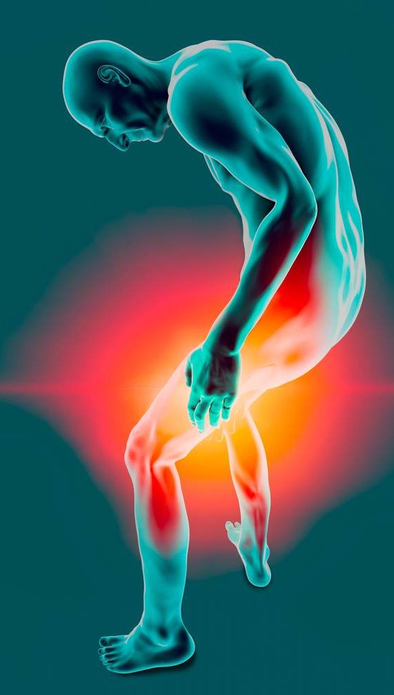
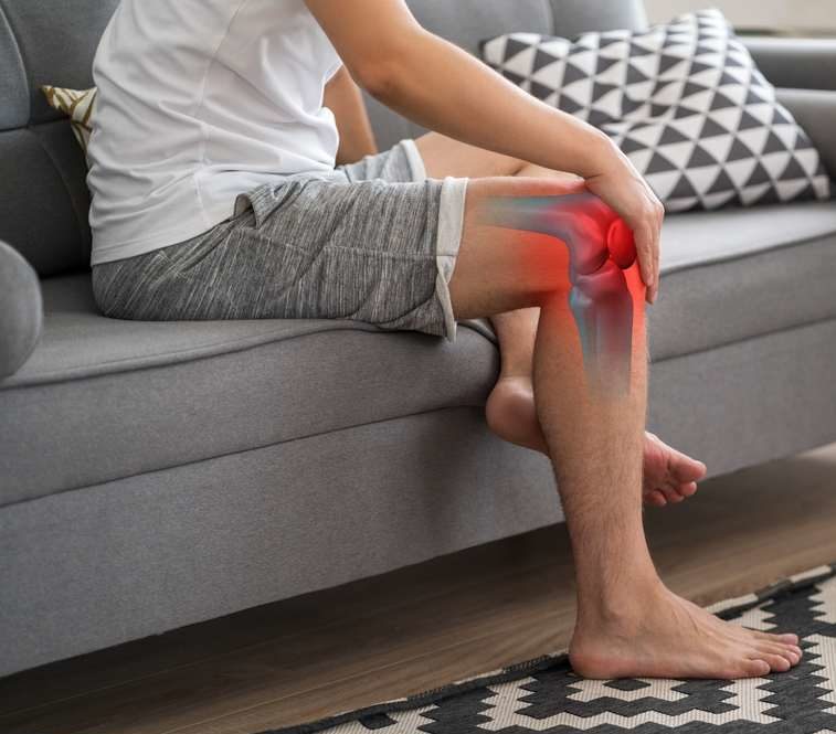
Signs and Symptoms of a Broken Femur
- Immediate pain in the hip region or thigh
- Visible disfiguration of the thigh and hip
- Boney fragments visible through the skin (if the fracture is open)
- Bruising in the hip or thigh
- Swelling in the hip, thigh, or lower leg
- Changes in the color and temperature of the leg and foot
- Inability to put weight bear or move the extremity
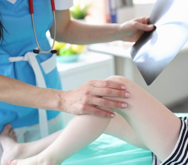
Diagnosis
As with any injury, the diagnosis of a broken leg starts with a history of what happened to cause the problem and an examination of the patient. Broken femurs are very painful and result in an inability to use the leg for walking, which means most people will be diagnosed in an acute care setting like the emergency department. The doctor will also assess you for any other injuries that may have occurred at the same time.
While the diagnosis of most femoral fractures can be made readily by simple radiographs (X-rays), the doctor may order more advanced imaging such as computed tomography (CT) scan or magnetic resonance imaging (MRI) to assess for soft tissue injuries that can complicate a broken femur. At some point, a bone density scan may be ordered to see if there is any osteoporosis in the other femur that may require treatment to prevent a fracture from occurring there in the future. If the femur was broken in a car accident, you will also likely get imaging to rule out other car accident injuries.
Types of Femoral Fractures
Open
Open fractures occur when the broken bone pierces the skin, leaving a wound in the tissue. This opening can allow bacteria to enter the wound and the bone fragments, which can lead to serious infections. An open femoral fracture requires repair not just of the fracture but of the soft tissues that have been damaged as well.
Closed
Closed fractures do not puncture the skin of the leg but can damage the surrounding soft tissue beneath the skin.
Stable
A stable fracture means the two parts of the broken femur still line up, making it much easier to treat.
Displaced
A displaced fracture means the two parts of the broken femur do not line up anymore, and so the treating physician must realign those fragments. This may require surgery and the placement of hardware to secure the bones in the correct place for healing.
Shaft Fractures
Shaft Fractures of the long straight center portion of the femur and include many different varieties of fractures.
Transverse fractures are straight across the width of the bone, going from side to side.
Oblique fractures are at an angle across the width of the bone, going from a high point on one side to a lower point on the other or vice versa.
Spiral fractures are associated with a twisting force that breaks the bone, resulting in a fracture that is highly oblique and spirals around the shaft of the bone, similar to a candy cane stripe.
Comminuted fractures occur when there are more than three fragments of bone left after the break.
Neck Fractures
Neck fractures are the most common type of broken femur to occur in those whose fractures are related to osteoporosis. These fractures are graded according to whether they are displaced or stable and to what extent, using the Garden Classification.
Distal Fractures
Distal fractures occur when the part of the femur that is furthest from the body and closest to the knee is broken. Due to its proximity to the knee, these fractures often include injury to that joint that will also require treatment.
How Do Broken Legs Heal?
When you break your femur, your body starts healing right away through a series of steps. First, a blood clot forms around the fracture to help protect and support the healing process. Over the next two to three weeks, your body creates a soft, cushion-like bridge made of tissue called a fibrocartilaginous callus. This acts like a temporary filler, holding the broken thigh bone pieces together while your body works on fixing the damage. As your femur continues to heal, the soft tissue gradually gets replaced by a stronger, mineral-rich bone callus. This stage, called ossification, can take one to four months. Even then, the bone isn’t fully healed.
If you’re wondering, “How long does it take for a broken femur to heal?” the answer is complicated. Since the femur is the longest and one of the largest bones in the body, the recovery time for a broken femur can be longer than for other broken bones. The healing process necessary strengthens the femur and helps restore its normal shape and function so that it’s as strong as it was before the injury.
Treatment Options for a Broken Femur
Because the femur is the longest and strongest bone in your body, a femur fracture requires medical intervention to ensure proper alignment and healing. Without properly stabilizing the bone, the powerful muscles surrounding the femur can pull the bone fragments out of place, which can lead to improper healing or even long-term complications or delayed recovery time for broken femur injuries.
Surgical Stabilization
In most cases, a femur fracture requires surgery to realign and secure the bone. The type of surgical intervention will depend on the location and severity of the break. Here are some examples of what that might be like:
- Internal Fixation: If the bone fragments need stabilization, the orthopedic surgeon may use metal plates, screws, rods, or nails to hold the pieces together. These devices are placed inside the leg, which means they are not visible externally.
- Intramedullary Nailing: A common technique for femur fractures, this procedure involves inserting a metal rod into the hollow center of the bone, known as the medullary canal, to keep it stable during healing. This surgical technique provides strong internal support and can even make for a faster recovery.
- External Fixation: In severe cases where immediate internal fixation is not possible, an external fixator may be used. This involves placing metal pins or screws into the bone above and below the fracture site, which are then connected to an external stabilizing frame outside the leg. This method is often used temporarily until further surgery can be performed.
Recovery and Rehabilitation
Healing from a femur fracture requires time, patience, and rehabilitation. After surgery, you may not be able to put weight on the injured leg for several weeks or even months, depending on the severity of the fracture and the type of surgical repair. Crutches, walkers, or wheelchairs are often necessary during this period. A broken thigh bone in elderly patients can also take longer to rehabilitate and requires consistent treatment.
Physical therapy also plays a crucial role in recovery, helping you regain strength, mobility, and flexibility. A rehabilitation plan typically includes:
- Range-of-motion exercises to prevent stiffness and maintain joint health
- Strength training to rebuild muscle support around the healing bone
- Gradual weight-bearing activities as the bone strengthens and becomes stable enough to support the body’s movement
Supporting Bone Healing
Recovering from a broken femur is a lengthy process, but with proper treatment, rehabilitation, and self-care, most patients regain full function and return to their normal activities. Several factors can influence how well and how quickly the femur heals from a fracture. To make the best of your recovery, here are some suggestions:
- Follow your doctor’s instructions carefully, including taking prescribed medications, attending follow-up appointments, and following all weight-bearing restrictions.
- Watch for signs of complications, such as increased pain, swelling, redness, or signs of infection, and report them to your doctor immediately.
- Avoid smoking and excessive alcohol consumption, since both can slow bone healing and increase the risk of complications.
Complications of a Broken Femur
Complications of a broken femur can be very mild to life-threatening, which makes it critical to discuss with your provider what risks you have personally and what signs and symptoms of these complications they want to be notified of immediately. Many factors influence the risk for such complications, such as how the leg was broken, age, weight, diet, medications, bone density, and more.
- Blood Loss can occur from the tearing of surrounding blood vessels and those that feed the bone itself. This blood loss can be severe, even in a closed fracture where the bleeding occurs into the tissue of the thigh. Damage to blood vessels can also lead to blood clots that can impair blood flow to the rest of the leg.
- Fat Embolisms happen when a tiny portion of the bone marrow, which is rich in fat, breaks off and enters the bloodstream. These little pieces of fatty marrow can then lodge in the lungs and cause a pulmonary embolism. While this is rare, it is a recognized complication of a broken femur.
- Shortened Limb is a risk of a severely comminuted fracture since aligning the pieces can sometimes result in a slight difference in the bone length after healing.
- Avascular Necrosis occurs when there is a lack of blood flow to the bone, causing it to become necrotic (dead). This is more common in fractures to the neck and head of the femur.
- Infection is a significant risk in patients who suffer an open fracture of the femur since the skin and tissue beneath are open to the environment. To reduce this risk, antibiotics are often part of the care plan for people with open fractures.
- Compartment Syndrome happens when the swelling in the muscles and other soft tissues of the thigh begins to compress blood vessels and nerves in the leg, preventing blood flow to the thigh and lower extremities. This pressure has to be relieved to save the tissue of the leg from harm.
- Nonunion is when two or more fragments of bone broken do not heal back together.
Broken Femur Care at AICA Orthopedics
The single best way to prevent complications of a broken femur is to seek out the care of an experienced and skilled provider who can ensure your care is managed appropriately. The doctors at AICA College Park are available to help you heal and recover, so get in touch today to schedule your appointment.